Home

ritm Administra Destul de lung markings stand frigider La bordul

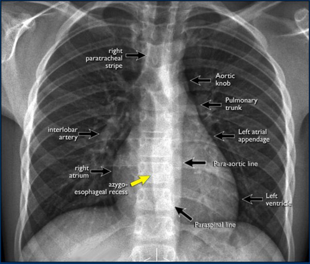
Chest radiograph - Wikipedia

Gopal Punjabi on Twitter: "PE on CXR. Note large R desc pulmonary artery  (Palla's sign), looks like sausage. Decreased vascular markings in R lung  (Westermark). 3D CT simulated XR, and CT image
Gopal Punjabi on Twitter: "PE on CXR. Note large R desc pulmonary artery (Palla's sign), looks like sausage. Decreased vascular markings in R lung (Westermark). 3D CT simulated XR, and CT image

Prominent Bronchovascular Markings in Chest X-Ray Report: All you need to  know - MyHealth
Prominent Bronchovascular Markings in Chest X-Ray Report: All you need to know - MyHealth

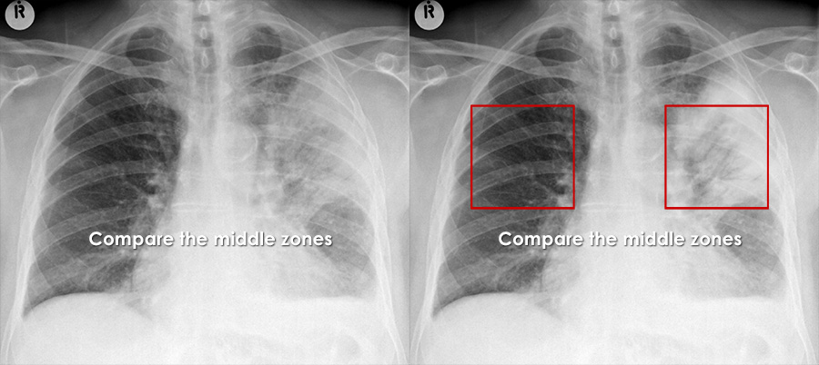
Interstitial Lung Disease or COVID-19? A Complex Pulmonary Differential  Amid a Global Health Crisis - Clinical Advisor
Interstitial Lung Disease or COVID-19? A Complex Pulmonary Differential Amid a Global Health Crisis - Clinical Advisor

EPOS™
EPOS™

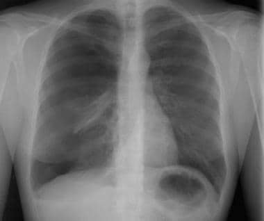
Normal Chest X-Ray and Lung - radRounds Radiology Network

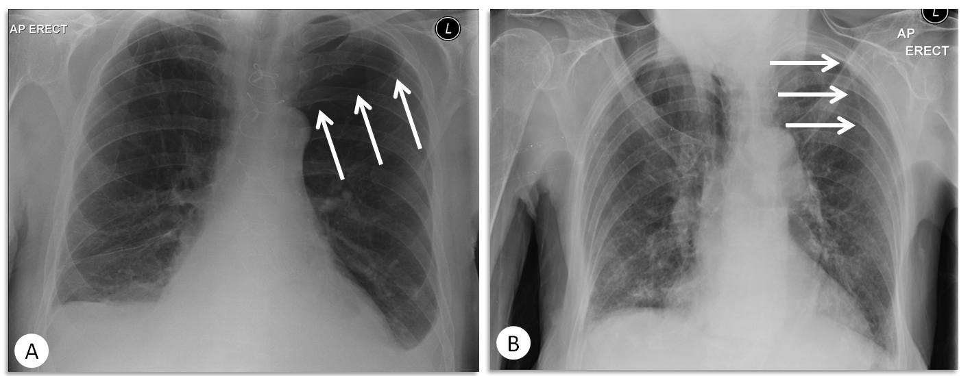
Pneumothorax Imaging: Practice Essentials, Radiography, Computed Tomography

ECMO‐dependent respiratory failure after snorting speed associated with  anti‐GBM antibodies - De Rosa - 2015 - Respirology Case Reports - Wiley  Online Library
ECMO‐dependent respiratory failure after snorting speed associated with anti‐GBM antibodies - De Rosa - 2015 - Respirology Case Reports - Wiley Online Library
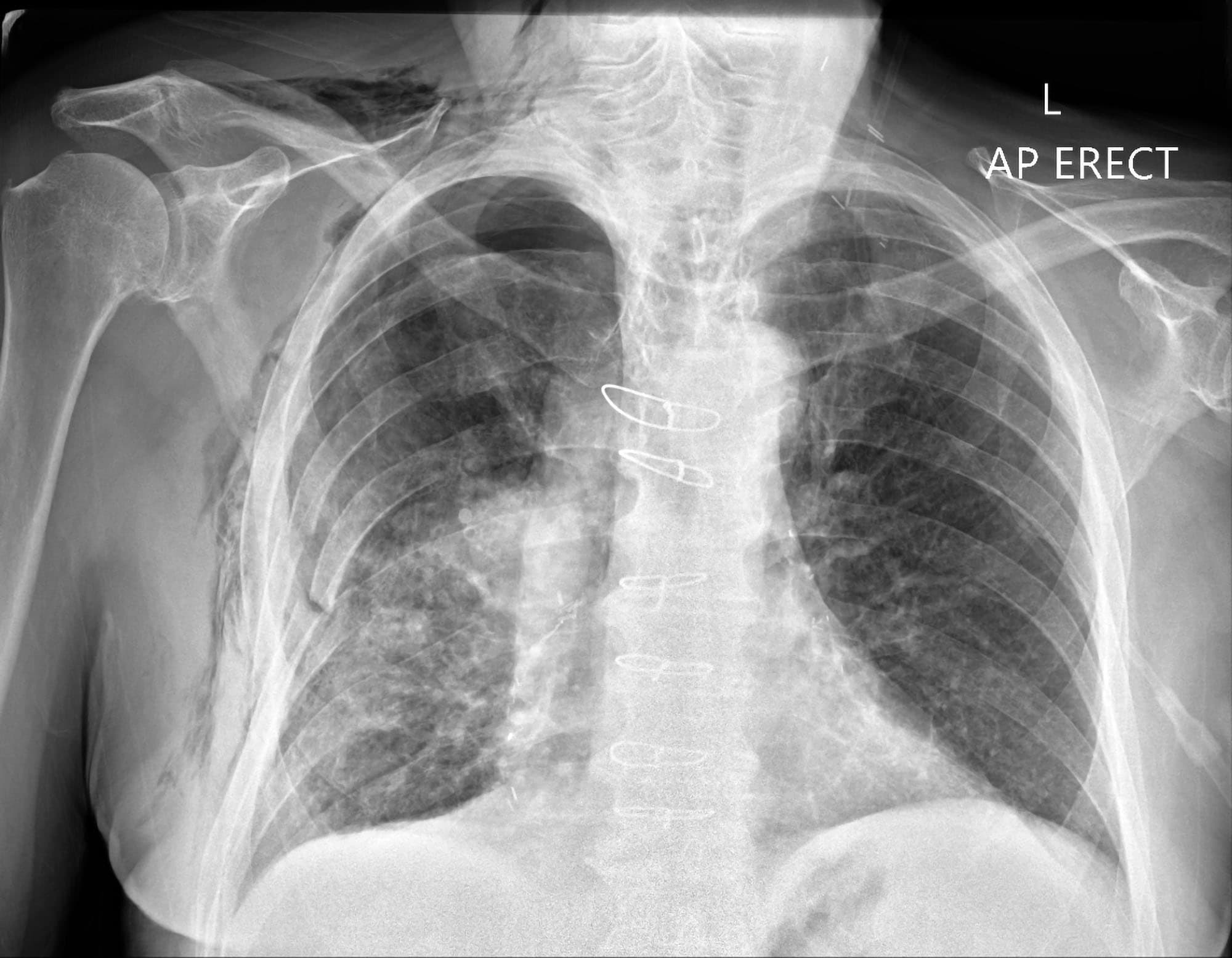
Chest X-ray Abnormalities - Lung abnormalities

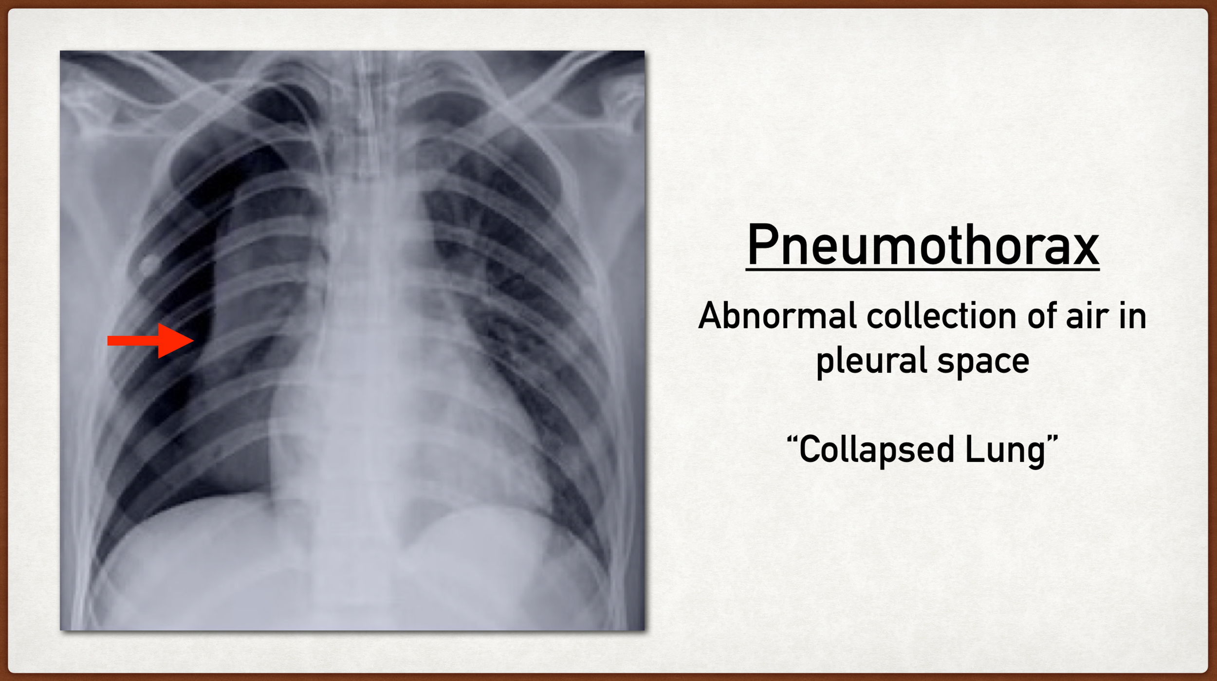
PT" Trick for Pneumothorax Types, Symptoms, Risk Factors, Causes, and  Treatment of Collapsed Lung — EZmed
PT" Trick for Pneumothorax Types, Symptoms, Risk Factors, Causes, and Treatment of Collapsed Lung — EZmed

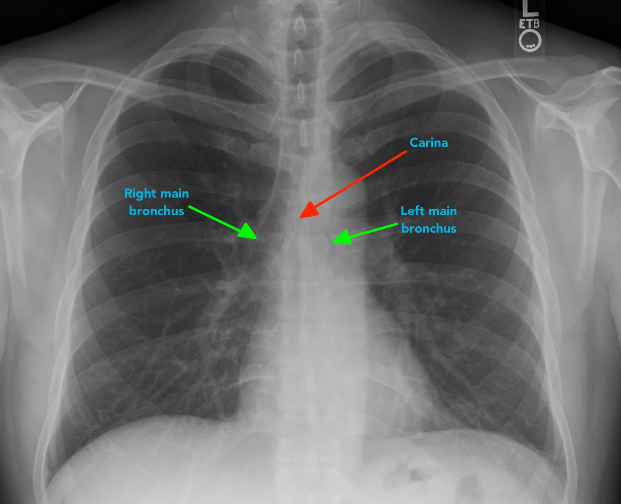
Chest X-ray Interpretation | A Structured Approach | Radiology | OSCE

COVID-19 pneumonia | Radiology Case | Radiopaedia.org

Medical Sciences, Pathology

Figure 1 | Thorax
Figure 1 | Thorax

Chest x-ray, Difference between Obstructive and Restrictive Lung Diseases -  YouTube
Chest x-ray, Difference between Obstructive and Restrictive Lung Diseases - YouTube

Chest Imaging Made Easy Course : lecture 03 ; part 1 Normal chest X-ray  Anatomy - YouTube
Chest Imaging Made Easy Course : lecture 03 ; part 1 Normal chest X-ray Anatomy - YouTube

View Image
View Image

What do increased lung markings in a chest x-ray mean? What are the most  likely causes of this? - Quora
What do increased lung markings in a chest x-ray mean? What are the most likely causes of this? - Quora

Hidoc Dr - Daily Medical Case Discussions - PULMONARY FIBROSIS. Increased,  coarse lung markings bilaterally mid & Lower zones more towards the  periphery of the lungs tracheal deviation towards RT side smaller
Hidoc Dr - Daily Medical Case Discussions - PULMONARY FIBROSIS. Increased, coarse lung markings bilaterally mid & Lower zones more towards the periphery of the lungs tracheal deviation towards RT side smaller

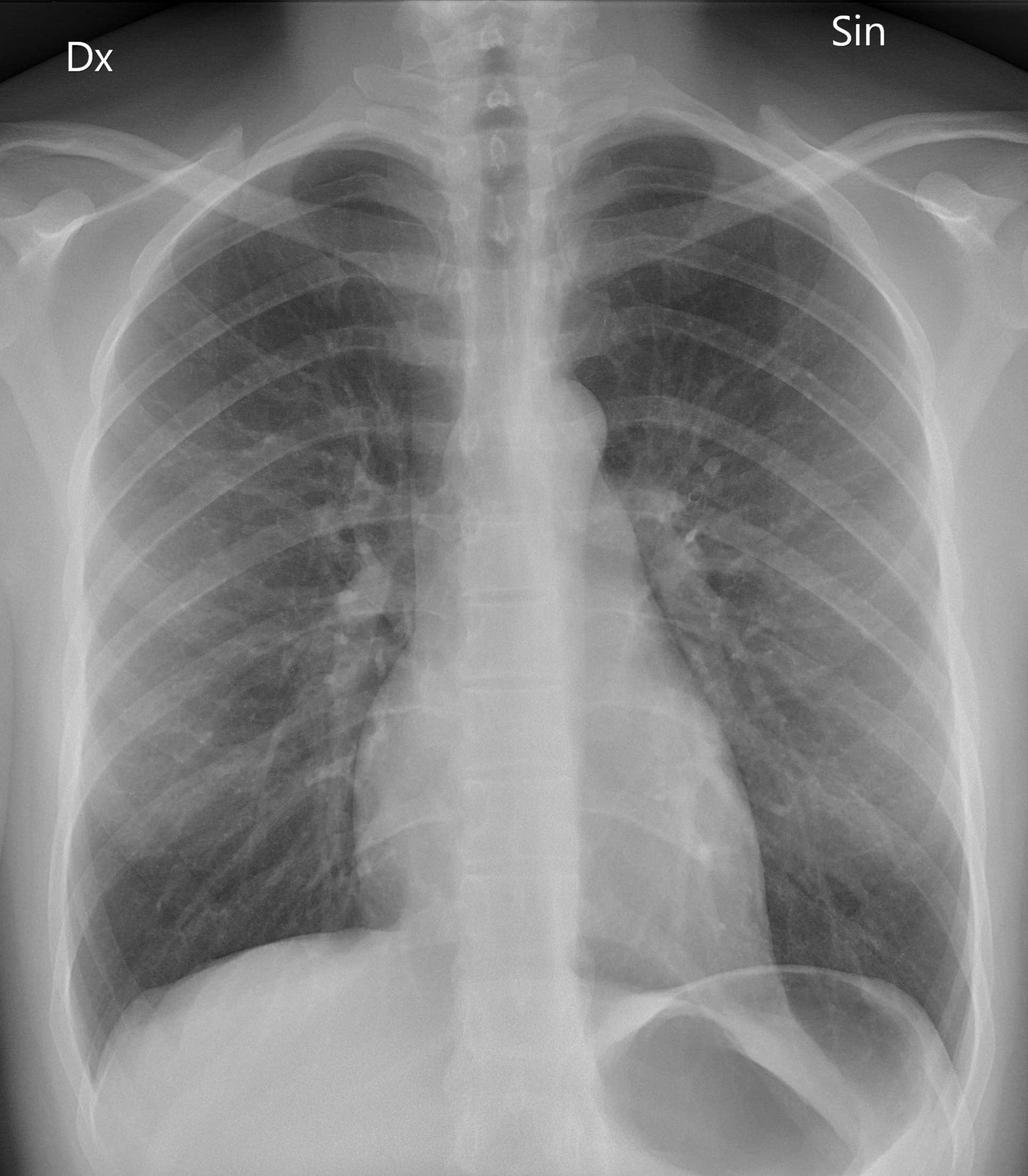
Patttern of lung disease on chest x ray

Radiology Rounds – 5/31/22 | PulmPEEPs

The Radiology Assistant : Chest X-Ray - Basic Interpretation

Loss of Lung Markings | Chest X-Ray - MedSchool

Chest radiograph showing increased lung markings in the right lower... |  Download Scientific Diagram
Chest radiograph showing increased lung markings in the right lower... | Download Scientific Diagram

Radiology In Ped Emerg Med, Vol 4, Case 5

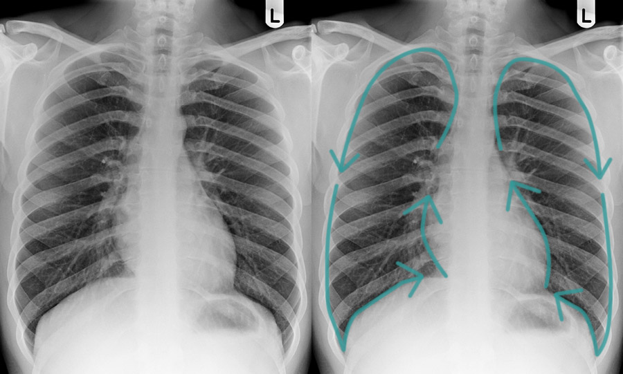
Chest X-ray Anatomy - Pleura and pleural spaces